


Foot & Ankle • Progressive Sports Medicine
Ankle Sprains & Syndesmosis Injuries
Most ankle sprains recover very well. The keys are recognising the ones that behave differently, especially high ankle (syndesmosis) injuries, and investing in the rehabilitation that prevents the next sprain.
Overview
Acute ankle sprains
Acute ankle sprains are common, can occur at any age, and generally recover very well. Most often, the ankle rolls inwards (called inversion) to cause these injuries, and most of the time you can take a few steps even though there is pain when putting weight on the ankle.
▶ Watch: Ankle sprains explained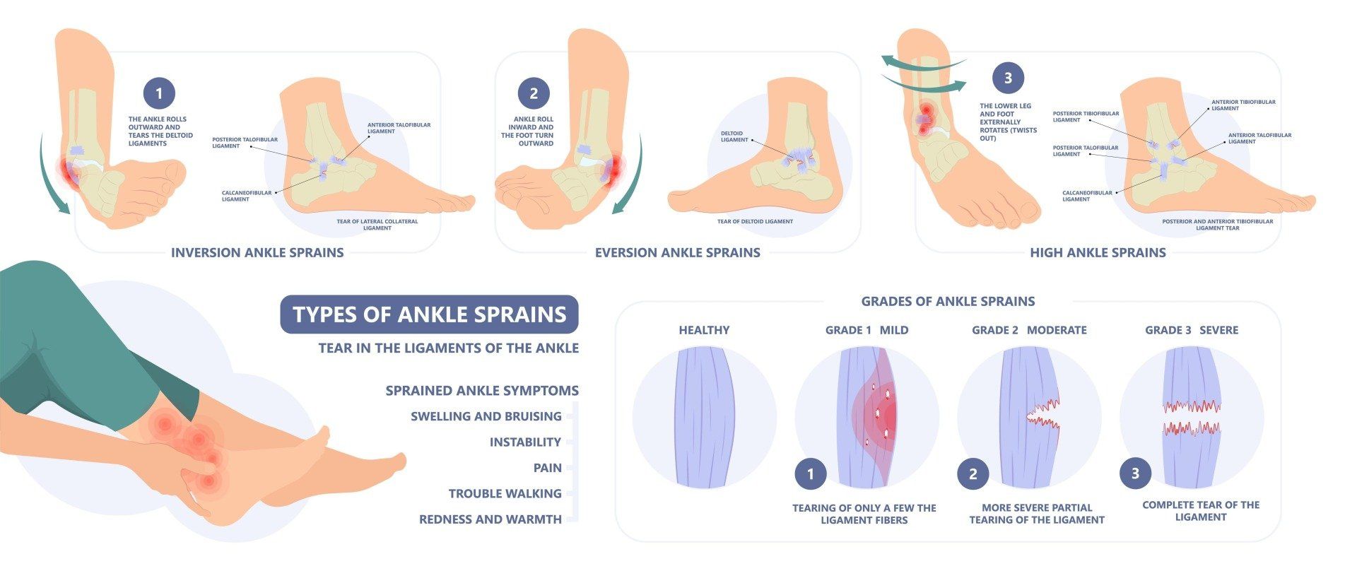
Severity
The three grades of ankle sprain
Grade 1
ATFL
The most common injury, generally involving only the ligament at the front, the anterior talofibular ligament. People generally recover very well and function even if this ligament does not heal fully.
Grade 2
+ CFL
Occurs as more force is put into that inverted position, partially or fully tearing the next ligament in the line, the calcaneofibular ligament (CFL).
Grade 3
Full tears
The highest grade, with full tears of both ligaments and sometimes injury to ligaments toward the back of the ankle.
Importantly, the evidence overall does not show a correlation between the grade of injury and long-term outcome.
Higher grade injuries may do better with early immobilisation, but only very short term, less than 10 days, because longer immobilisation can lead to increased stiffness.
We know there is an increased risk of re-injuring the same ankle, because the torn ligaments serve proprioceptive functions (your sense of joint position). This is the main value in ensuring patients invest in a proper course of rehabilitation: future injury prevention.
When sprains recur
Chronic ankle instability
Unfortunately, some patients progress to recurrent ankle sprains, which are frustrating and may predispose to ankle joint damage.
Instability is something that only a patient can tell us. It cannot be concluded simply by examination, which is why a doctor needs to really listen.
A patient who does not trust their ankle, is worried about walking in the dark, and has usually had a number of sprains, has chronic ankle instability. They may also have pain between episodes, and most have had to modify their lifestyle and activities around the injury.
The most common cause is inadequate rehabilitation. Most clinicians do not refer patients without other serious problems for surgery until they have trialled good quality rehabilitation for at least 6 weeks, in combination with bracing and taping to assist proprioception.
Evidence shows that taping and bracing reduce the risk of recurrent sprain immediately, to the same level that rehabilitation alone would reach by 10 weeks.
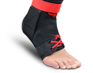
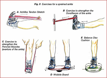
If, despite this, there is ongoing ankle pain, swelling and more than 3 episodes of significant ankle sprain in a year, then a patient with chronic ankle instability really should see a foot and ankle surgeon for an opinion.
High ankle injuries
Ankle syndesmosis injuries
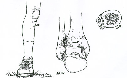
Be alert for this pattern: an external “foot out” rotational mechanism, generalised swelling, and pain higher up on the ankle. This could be a syndesmosis or high ankle injury, which may be unstable and should be referred for further workup.
Most of the time it’s very hard to remember which way the foot went, so when there is pain higher than expected we need to be on alert for this potential injury.
The ankle joint normally functions like a hinge, with the two lower bones of the leg (the tibia and fibula) locking into the saddle-shaped bone in the middle, called the talus. When a forceful sideways motion occurs, it opens the hinge and spreads the strong ligaments that hold the two bones together. This always occurs in a front-to-back pattern, like a book opening up, so tenderness around this area of the ankle is quite suggestive of injury.
▶ Watch: How a syndesmosis injury happens
Diagnosis & treatment
Diagnosing and treating syndesmosis injuries
Making the diagnosis
The diagnosis can be made quite confidently through a combination of clinical tests. An ultrasound adds the advantage of being able to see the ligament at the front for any damage, and MRI can determine whether there are any associated injuries inside the ankle joint.
First principles
As with any acute injury, the principles are to control the initial inflammatory response with rest and optimal loading, ice, compression and elevation. The severity of ligament damage, or associated cartilage or bony injury, determines whether surgery is needed.
Most cases are lower grade injuries that can certainly be managed non-operatively, following the milestones below, with the use of a stirrup that helps push the ends of the bones together, so the ligament heals in a tighter, more functional position.
3 weeks sooner
Our research: PRP for syndesmosis injuries
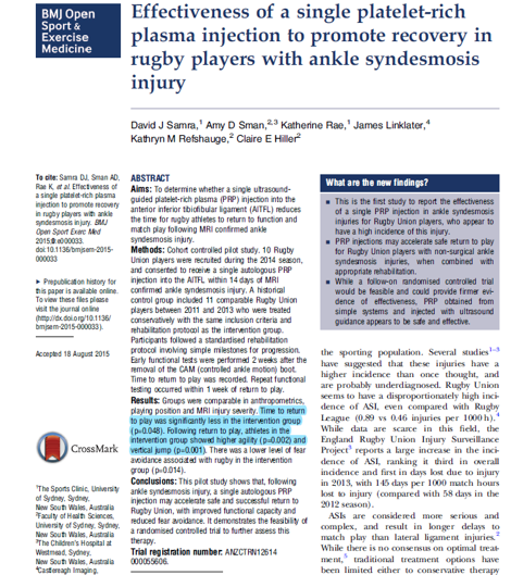
In research led by Dr Samra, rugby players with ankle syndesmosis injuries returned to play an average of 3 weeks sooner with a single platelet-rich plasma (PRP) injection to the torn ligament, in combination with standard rehabilitation.
Samra DJ, et al. Effectiveness of a single platelet-rich plasma injection to promote recovery in rugby players with ankle syndesmosis injury. BMJ Open Sport & Exercise Medicine 2015;1:e000033. doi:10.1136/bmjsem-2015-000033
Recovery
Rehabilitation milestones
For stable syndesmosis injuries managed non-operatively, progression is guided by pain-free functional milestones rather than the calendar:
- 1
Surgery if unstable. Stable injuries can expect a 6–10 week recovery.
- 2
A single platelet-rich plasma injection may accelerate recovery (see the research above).
- 3
Ankle stirrup (or boot) and non-weight bearing until a pain-free double leg lunge.
- 4
Stirrup and walking only until a pain-free single leg lunge.
- 5
Stirrup and cross training(bike, pool, Alter-G) until pain-free hopping.
- 6
Stirrup and straight line running.
- 7
Sport-specific rehabilitation once pain-free running for 1 week.
- 8
The stirrup should be weaned gradually.
Common questions
Ankle & syndesmosis FAQs
The clues are the mechanism and the location of pain. A forceful external ’foot out’ rotation, generalised swelling, and pain higher up on the ankle than a typical sprain all raise suspicion of a syndesmosis injury. Most people can’t remember which way the foot went, so pain higher than expected is itself a reason to be assessed, because unstable syndesmosis injuries need further workup.
It depends on the assessment. The diagnosis can often be made confidently through clinical tests. Ultrasound adds the ability to see the ligament at the front of the syndesmosis for damage, and MRI can determine whether there are associated injuries inside the ankle joint, which influence treatment.
Simple ankle sprains generally recover well over days to weeks with optimal loading. A stable syndesmosis injury typically takes 6 to 10 weeks, progressing through a structured milestone program in an ankle stirrup. Unstable syndesmosis injuries require surgery, which lengthens the timeline.
For ankle syndesmosis injuries, research led by Dr Samra found that rugby players returned to play an average of 3 weeks sooner with a single platelet-rich plasma injection to the torn ligament, in combination with standard rehabilitation. Whether PRP is appropriate for your injury is determined at assessment.
The torn ligaments serve proprioceptive functions, so re-injury risk rises after a sprain, and inadequate rehabilitation is the most common cause of recurrence. Good quality rehabilitation for at least 6 weeks with bracing or taping is first line. If there is still ongoing pain, swelling and more than 3 significant sprains in a year, an opinion from a foot and ankle surgeon is appropriate.
Rolled your ankle, or pain higher than a normal sprain?
Our Sport and Exercise Physicians in Leichhardt can assess your ankle, use point-of-care ultrasound to examine the ligaments, and build a milestone-based plan, informed by our own published research on syndesmosis injuries.
This page provides general information only and is not a substitute for personalised medical advice. Please consult a qualified health practitioner for assessment of your individual circumstances.