


Shoulder • Progressive Sports Medicine
Rotator Cuff Syndrome: Impingement, Bursitis & Tears
From swimmer’s shoulder to rotator cuff tears, these conditions sit on one spectrum. Understanding where you are on it, and fixing the mechanics behind it, is what gets shoulders better.
Introduction
One spectrum, from irritation to tears
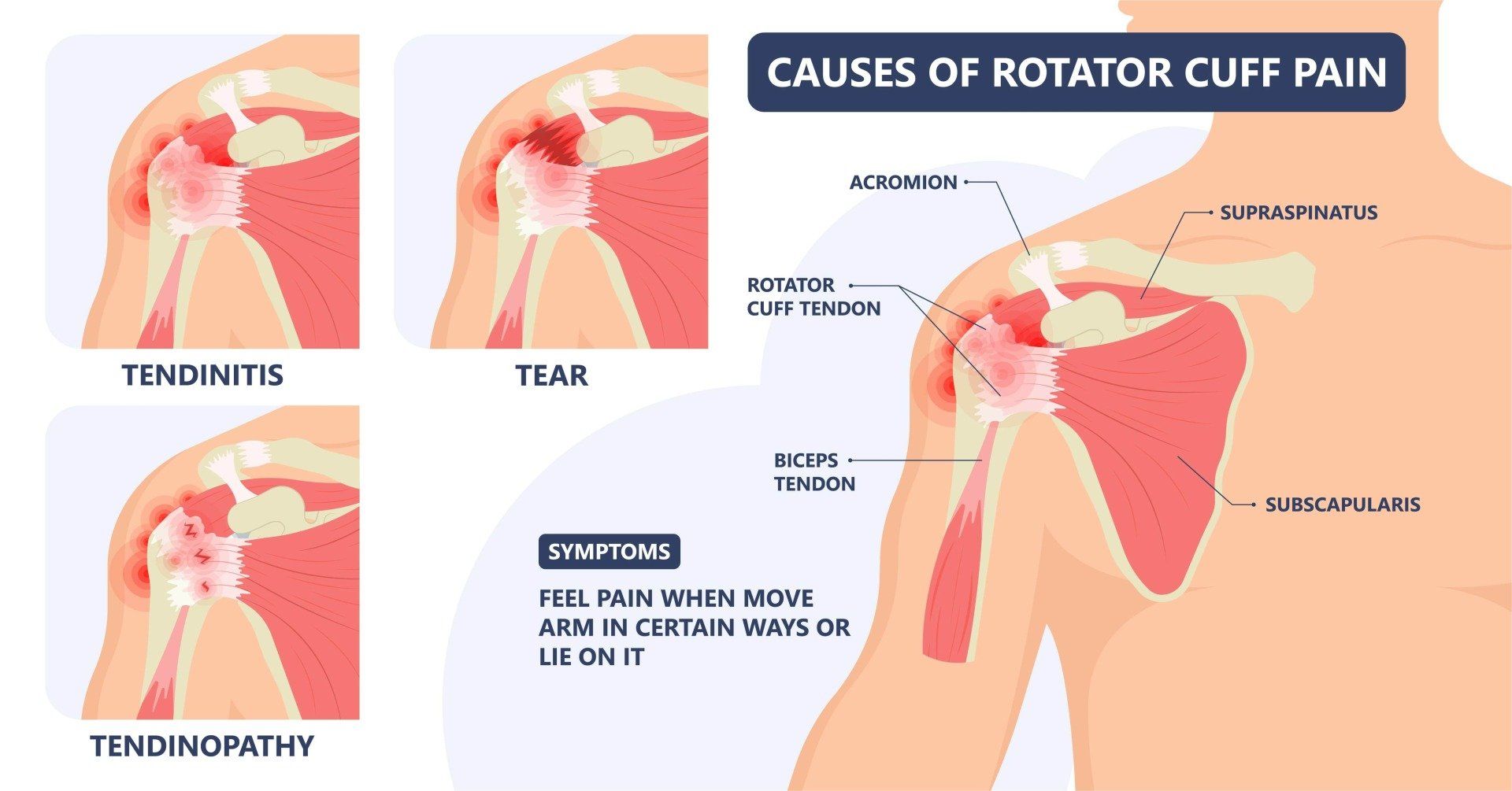
Swimmer’s Shoulder is a condition that covers a broad spectrum of pathology. Due to repetitive overhead activity, there is mechanical irritation of the tissues under the arch of the shoulder. As it progresses, the rotator cuff tendon becomes irritated and then develops features of disrepair. When microscopic tears come together over a long period, and the rate of tissue breakdown slowly exceeds the rate of repair, the condition transitions along the spectrum:
Stage 1
Bursitis & impingement
Mechanical irritation of the bursa and tissues under the bony arch of the shoulder.
Stage 2
Tendinopathy
The rotator cuff tendon becomes irritated and develops features of disrepair.
Stage 3
Cuff tears
Accumulated microscopic tears join as breakdown outpaces repair, usually as partial tears from wear.
The mechanism
Subacromial bursitis and shoulder impingement
The shoulder is a complex joint where several bones, muscles and ligaments connect the upper limb to the chest. The subacromial space is the area underneath the bony arch of the shoulder, and like many areas of tissue movement, it has a bursa: a thin, lubricating fluid sac that helps tissue planes glide.
In the video below you can see an anatomical section of the shoulder with the ball and socket, the rotator cuff, and this layer of tissue that helps it slide under the bony arch as you lift your arm.
 Inside the shoulder: the bursa & rotator cuff ▶ Watch on YouTube
Just like most forms of deep bursitis, it often coexists with tendon irritation and wear. When the bursa becomes inflamed it swells and causes pain. Inflammation leads to swelling and thickening of the tendons and bursa, reducing the space and further squeezing the rotator cuff tendon and bursa. More importantly, pain leads to maladaptive movement of the shoulder: imbalances in muscle forces push the ball upward in the socket, again pinching the tendons under the bone. The cycle continues, and this compression results in pain, tenderness and sometimes weakness.
The combined condition of rotator cuff tendinosis and bursitis is called an impingement syndrome. It can be structural (a bony spur or narrowing under the acromion) or functional (poor rotator cuff function and force imbalances).
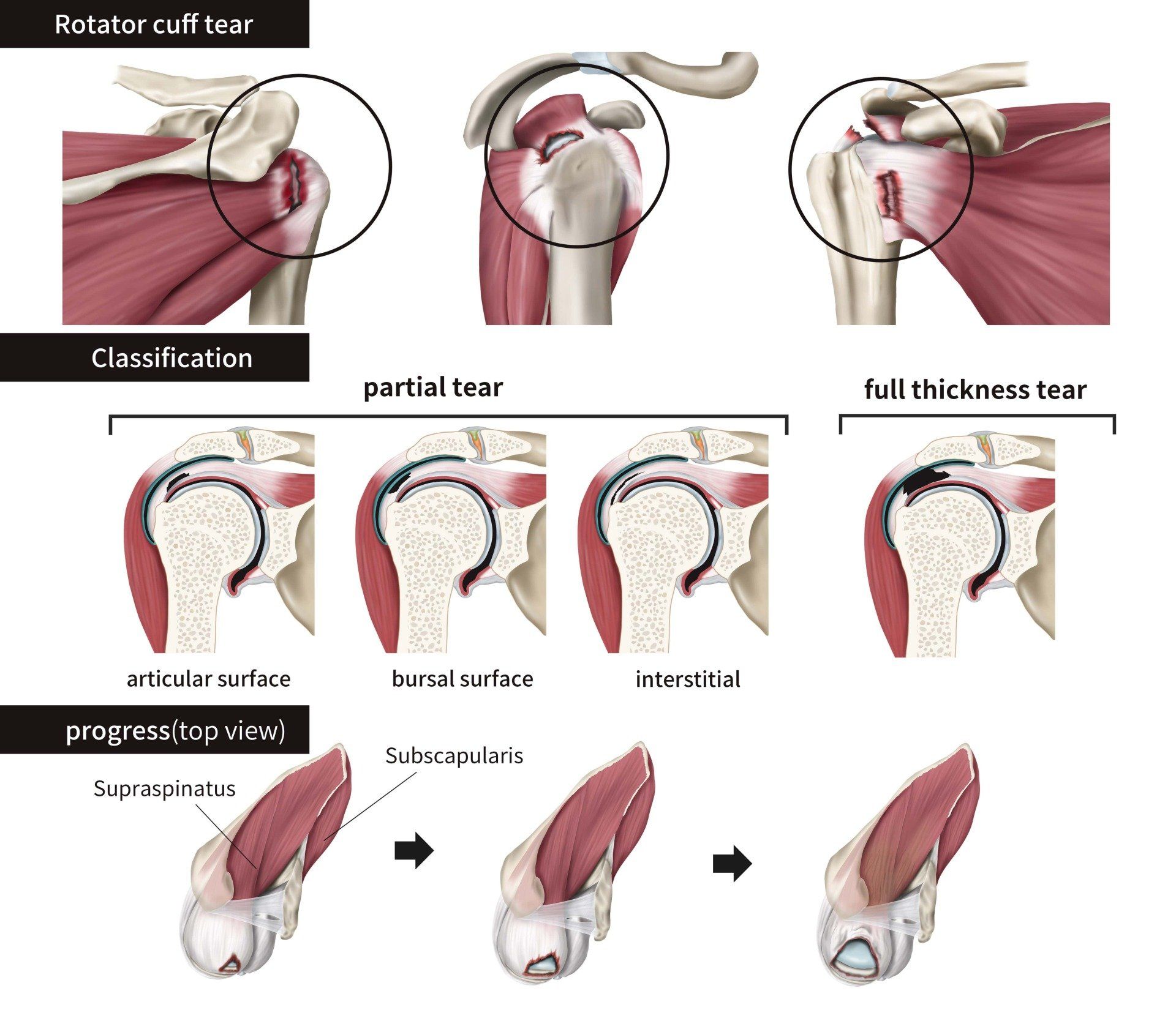
With age and continued repetitive use, rotator cuff tears can occur. When these happen without trauma, they are usually due to wear. Most involve the top part, the supraspinatus tendon, and most are partial tears(the ends together but frayed) rather than full-thickness tears (the ends separated). When there is a big full-thickness tear there is usually weakness, and most patients require surgery to restore their strength.
Causes
Why swimmer’s shoulder develops
Swimmer’s shoulder and bursitis may be caused by repeated minor trauma, such as overuse of the shoulder joint and muscles during swimming, but also other activities like overhead weights, throwing, and even occupational and domestic tasks.
Despite most swimmers being very well trained, the sheer number of repetitive cycles of swimming is the major factor. Under fatigue, the muscles that control shoulder movement can fail to clear the space for the rotator cuff and bursa, so impingement occurs.
Diagnosis & treatment
Diagnosing and treating subacromial bursitis
The medical workup involves a comprehensive history, physical examination, and often point-of-care dynamic ultrasound to assess the movement of tissues as they glide under the acromion.
Bursitis is a bit like smoke to fire in most cases. The bursitis is a downstream effect of abnormal shoulder mechanics: pain inhibits normal muscle activation, which perpetuates those mechanics, and usually goes with irritation of the rotator cuff itself. This requires a holistic view of treatment.
First line treatment for bursitis should be:
- Avoid the triggers for irritation: overhead activities, lifting, and anything that provokes pain.
- X-rays to exclude a primary, structural source of impingement.
- NSAIDs at anti-inflammatory (not just pain-relieving) doses, for 7–10 days.
- Rehabilitation exercises to restore balance, centring of the humeral head, and stability of the base of the shoulder.
- Escalation to a corticosteroid injection , a more potent anti-inflammatory, if you are unable to make progress. Injections are delivered under ultrasound guidance for accuracy.

An injection needs to be combined with rehabilitation exercises about one week afterwards, so that pain is no longer inhibiting proper muscle activation. The results of surgical decompression are controversial, with studies showing no long-term benefit of either open or arthroscopic debridement compared to an exercise-based approach at 2 years.
If you have shoulder pain and impingement, see a health professional with expertise to get a diagnosis and a treatment plan.
When the tendon tears
Rotator cuff tears
The rotator cuff is a broad, flat group of tendons that come from the wing bone (scapula) to wrap around the ball of the shoulder, which doctors call the head of the humerus. They pull the ball into its socket and keep it centred with all movements of the shoulder. The cuff also helps us elevate the arm overhead, and this movement is lost when the rotator cuff is not working.
When a patient presents with a painful, weak shoulder, the diagnosis of a rotator cuff tear is best confirmed with MRI in corroboration with clinical examination findings, picking up around 85% of tears. Ultrasound is useful in experienced hands and can be corroborated with the examination immediately.
Full-thickness tears are any tear that goes from top to bottom through the rotator cuff. They can be wide or small, and we also measure the degree of retraction: how far the tendon ends have moved away from each other. As you can see in the video below, sutures with anchors that invest the tendon back to its bony footprint are used to repair a full-thickness tear.
 How a rotator cuff repair works ▶ Watch on YouTube
Common questions
Rotator cuff & impingement FAQs
Usually not on its own. Bursitis is a bit like smoke to fire: it is a downstream effect of abnormal shoulder mechanics. Pain inhibits normal muscle activation, which perpetuates those mechanics and irritates the rotator cuff itself, so treatment has to take a holistic view rather than only settling the bursa.
Rarely. The results of surgical decompression are controversial, with studies showing no long-term benefit of either open or arthroscopic debridement compared with an exercise-based approach at 2 years. Rehabilitation that restores balance and centring of the ball in the socket is the foundation of treatment.
It depends on the type. Most rotator cuff tears are partial tears that happen over time due to wear, and these are generally managed without surgery. A large full-thickness tear, where the tendon ends are separated, usually causes weakness, and most patients in that situation require surgical repair to restore strength.
As an escalation, not a starting point: if triggers have been avoided, anti-inflammatories trialled and rehabilitation is unable to progress because of pain, a corticosteroid injection can settle the inflammation. It needs to be combined with rehabilitation exercises about one week after the injection, so the pain is no longer inhibiting proper muscle activation.
Through a comprehensive history and physical examination, often with point-of-care dynamic ultrasound to watch the tissues glide under the acromion in real time. For suspected cuff tears, MRI corroborated with examination findings picks up around 85% of tears, and ultrasound is useful in experienced hands with immediate correlation to the examination.
A shoulder that pinches, aches at night, or won’t lift?
Our Sport and Exercise Physicians in Leichhardt can assess your shoulder with dynamic point-of-care ultrasound, work out where you sit on the spectrum, and build a plan that fixes the mechanics, not just the smoke.
This page provides general information only and is not a substitute for personalised medical advice. Please consult a qualified health practitioner for assessment of your individual circumstances.