


Heel & Foot • Progressive Sports Medicine
Plantar Fasciitis
The most common cause of heel pain, and one of the most misunderstood. It is not simple inflammation, it is rarely just the foot, and it almost never needs surgery. Getting it right means finding what is overloading the fascia, in your mechanics and in your metabolic health, and rebuilding its capacity.
Introduction
What is plantar fasciitis?
Plantar fasciitis is the most common cause of pain under the heel. This area of the body takes an enormous amount of both tensile and compressive load, so it doesn’t take much to develop problems here.
Plantar fasciitis is more correctly called plantar fasciosis or fasciopathy, because it is not a classically inflammatory problem, even if some inflammatory chemicals are present.
It is a very common presentation to Sports Physicians because surgery is an absolute last resort, and conservative management and time are usually effective. The duration of symptoms can be anywhere from months to years, but most cases resolve with treatment.
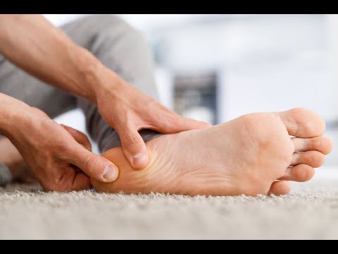 Watch: the anatomy of heel pain ▶ Watch on YouTube
Anatomy
The plantar fascia and the windlass
The plantar fascia (plantar aponeurosis) is a thick band of connective tissue that spans the sole of the foot, from the heel bone forward to the base of the toes. It is a key part of the arch’s support system and a spring that stores and returns energy with every step.

Its behaviour is governed by the windlass mechanism: as the big toe extends during push-off, the fascia winds around the joint like a rope on a drum, tightening the arch and stiffening the foot into a rigid lever. This is what a positive Windlass test reproduces in the clinic, and it is central to how the fascia is loaded and to how we treat it.


The two drivers
What drives plantar fasciopathy
Two forces combine to overload the fascia: the mechanical load going through it, and the quality of the tissue taking that load. Both are treatable, and both are worth understanding.
1. Mechanical overload
Sudden increases in standing, walking or running, tight calves that transmit extra tension to the heel, footwear, body weight and foot mechanics all raise the load through the fascia. The common risk factors are well recognised.

Body weight is a major and modifiable contributor, both through raw load and through its effect on metabolic health, which brings us to the second driver.

2. Tissue quality and metabolic health
The fascia is collagen, and collagen quality is not fixed. In diabetes and poor metabolic health, sugar molecules bind to collagen in a process called glycation, forming advanced glycation end-products that cross-link and stiffen the collagen, disorganise its structure and thicken the fascia. This is part of why heel pain is more common and more stubborn in people with metabolic disease.

This is why a stubborn plantar fasciopathy is sometimes a prompt to look wider. Assessing and improving metabolic health is not a distraction from treating the heel; for some patients it is part of treating the heel.
Diagnosis
Getting the diagnosis right
Plantar fasciopathy is largely a clinical diagnosis, from the history (classic first-step pain in the morning) and examination (tenderness at the fascia’s attachment, a positive windlass test). But heel pain has important mimics, and part of a thorough assessment is excluding them.
Other causes of heel pain we look for: a stress fracture or bone bruising of the heel bone, entrapment of the inferior branch of the lateral plantar nerve (which tends to create more burning pain), and bruising or inflammation of the fat pad underneath the heel. Each is managed differently, which is why the diagnosis comes first.
Point-of-care ultrasound is an extension of the examination: it shows thickening of the fascia at the heel and any changes within it, and helps distinguish fasciopathy from the mimics above.

Treatment
Treatment: reduce the load, rebuild the tissue
Treatment follows a logical ladder, and the great majority of people never need to reach the top of it. The foundations, done well and patiently, resolve most cases.
Settle the load. Relative rest from the aggravating activities, calf and fascia stretching, supportive footwear, orthotics or heel cups to offload the fascia, night splints to hold it out of its shortened position, and taping. Maintaining fitness through low-impact options such as cycling protects your conditioning while the fascia settles.


Rebuild the tissue. The fascia responds to load the same way tendons do. Progressive high-load strength training, such as heel raises performed with the toes propped up on a rolled towel to engage the windlass, has been shown to improve outcomes, and sits at the heart of modern rehabilitation. You can read more about how loading remodels this kind of tissue on our tendon pain page, and progress is supervised by our exercise physiology team.
Adjuncts for stubborn cases. When progress stalls, shockwave therapy can desensitise the fascia and stimulate a healing response, and is well supported by evidence in chronic plantar fasciopathy.

Where pain is blocking rehabilitation, a targeted injection can help, delivered under ultrasound guidance for accuracy and to protect the fat pad. We are judicious with corticosteroid at the heel, because repeated injections risk fat pad atrophy and fascial rupture, so it is used sparingly and precisely.

Surgery is an absolute last resort. With an accurate diagnosis, attention to both the load and the tissue, and patience, the great majority of plantar fasciopathy resolves without it.
The evidence
Key references
- Gautieri A, et al. Advanced glycation end-products: mechanics of aged collagen from molecule to tissue. Matrix Biol 2017;59:95–108. doi:10.1016/j.matbio.2016.09.001
- De Garceau D, et al. The association between diagnosis of plantar fasciitis and Windlass test results. Foot Ankle Int 2003;24:251–255. doi:10.1177/107110070302400309
- Rathleff MS, et al. High-load strength training improves outcome in patients with plantar fasciitis: a randomized controlled trial with 12-month follow-up. Scand J Med Sci Sports 2015;25:e292–e300. doi:10.1111/sms.12313
Common questions
Plantar fasciitis FAQs
Correct: plantar fasciitis is more accurately called plantar fasciosis or fasciopathy, because it is not a classically inflammatory problem, even if some inflammatory chemicals are present. It is a degenerative overload of the fascia. That distinction matters, because it points treatment toward restoring the tissue’s capacity through loading, rather than simply chasing inflammation.
Overnight the fascia rests in a shortened position, and the first loaded steps of the day suddenly stretch and compress a tissue that has lost some of its tolerance. That first-step pain, easing as you warm up and then returning after rest, is the classic pattern. It is also why the first treatments target that morning load, and why we look at what is driving the overload in the first place.
Yes, and sorting that out is the point of a proper assessment. Other important problems include a stress fracture or bone bruising of the heel bone, entrapment of the inferior branch of the lateral plantar nerve (which tends to create more burning pain), and bruising or inflammation of the fat pad underneath the heel. Each is managed differently, so an accurate diagnosis comes first.
More than most people realise. The fascia is collagen, and collagen is affected by metabolic health: in diabetes and poor metabolic health, sugar molecules bind to collagen (glycation), stiffening and disorganising it and thickening the fascia. This is one reason heel pain can be stubborn, and why we look at the whole person, not just the foot.
Almost certainly not. Surgery is an absolute last resort. Conservative management and time are usually effective: the duration of symptoms can be anywhere from months to years, but most cases resolve with treatment. The work is in identifying and correcting what is overloading the fascia, then progressively rebuilding its capacity.
Heel pain that bites with your first steps every morning?
Our Sport and Exercise Physicians in Leichhardt can confirm the diagnosis, rule out the mimics with point-of-care ultrasound, look at both the load and your metabolic health, and build the plan that actually resolves it.
This page provides general information only and is not a substitute for personalised medical advice. Please consult a qualified health practitioner for assessment of your individual circumstances.