


Knee Conditions • Progressive Sports Medicine
Jumper’s Knee (Patellar Tendinopathy)
Focal pain just below the kneecap that flares with jumping, landing, stairs and squatting. With the right diagnosis and a graded loading program, recovery is slow but sure.
Introduction
What is Jumper’s Knee?
Jumper’s Knee is similar to “Runner’s Knee”. Both are common terms often used to describe pain at the front of the knee. Jumper’s Knee refers specifically to a problem in the patellar tendon. These problems and pain can be related to:
Thickening & wear
Structural change within the tendon, known as patellar tendinosis.
Accumulated micro-tears
Tiny tears that can accumulate to form a visible tear, more akin to wear than a sudden injury.
Inflammatory chemicals
Irritant chemical signalling within the tendon. Notably, there are usually no inflammatory cells in tendons.
As the name suggests, Jumper’s Knee is a common complaint among athletes and people involved in plyometric sports such as netball, basketball and volleyball.
Anatomy
Where the pain comes from
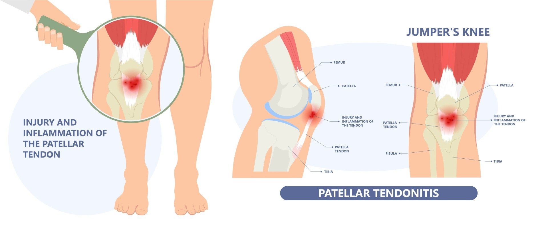
The pain usually occurs under or around the front of the kneecap (patella). You can usually point to the area with one finger.
The patellar tendon begins (or originates) on the patella and travels down the front of the knee to insert on the top of the shin bone, at a point called the tibial tubercle. When the thigh’s quadriceps muscles contract, the patellar tendon is pulled, which in turn straightens the knee and extends the leg.
The kneecap or patella is a sesamoid bone that is embedded in a tendon connecting the muscles of the thigh to the shin bone (tibia). The function of the patella is to protect the front part of the knee.
Causes
Why Jumper’s Knee develops
The patellar tendon moves every time the knee bends or straightens. Over time, overuse of the knee can cause tiny tears to form within the patellar tendon, which causes Jumper’s Knee. For example, a basketball player who jumps up and down on a parquet floor every day may experience mini-traumas to the patellar tendon that eventually cause painful knee symptoms.
Typically, Jumper’s Knee is caused by one or all of these factors:
Overuse or sudden stress
Sudden overload of the patellar tendon (such as a heavy or awkward landing) can cause inflammation or tears in the tendon tissue, with pain, swelling and discomfort during jumping, running and other athletic activities.
Deconditioning
Excessive or sudden rest, often due to illness or a separate injury, leaves the tendon less able to tolerate a return to normal load.
Lower limb rigidity
Stiffness of other joints, such as the hips, means they contribute less to shock absorption, transferring extra load to the patellar tendon.
These factors can cause the tendon to progress along the “continuum” of tendinopathy, leading to more degenerative changes that are very slow to remodel. With this in mind, athletes who find that the initial symptoms of Jumper’s Knee are not easing with a few days of rest should seek a medical evaluation to determine the best ways to prevent further injury.
Types
The continuum of patellar tendinopathy
Jumper’s Knee can be classified three ways, based on a continuum of pathology:
Stage 1
Reactive
Acute Jumper’s Knee, occurring from sudden, unexpected overexertion of the patellar tendon.
Stage 2
Disrepair
Continued symptoms that require careful activity modification and load management.
Stage 3
Degeneration
Chronic Jumper’s Knee, which manifests as a recurring problem over time.
Symptoms
What Jumper’s Knee feels like
The most common symptom is a dull aching pain underneath the kneecap while walking up or down stairs, squatting, kneeling, and sitting with your knees bent for a long period of time. Individuals with patellar tendinopathy may experience some or all of the following:
Pain during athletic motion
A sharp, throbbing pain beneath the kneecap during a workout or competition. At first the pain may worsen with activity and recede with rest. Left untreated, it may become constant, present even at rest, and worst when kicking, running or bending the knee, as these actions load the patellar tendon.
Swelling
Like most patellar injuries, Jumper’s Knee may cause mild swelling of the knee joint, and the knee may look swollen with a reduced range of motion.
Discomfort during daily activities
The patellar tendon helps extend the knee during everyday tasks such as kicking, climbing stairs or bending down. Advanced cases can cause increased pain during these daily activities.
Focal tenderness
Tenderness concentrated at one small point just below the kneecap, rather than spread across the front of the knee.
The hallmark of patellar tendinopathy that distinguishes it from runner’s knee is focal, direct tenderness under the kneecap the size of one or two fingers.
Diagnosis
How we diagnose Jumper’s Knee
Patients who suspect they have sustained a knee injury should seek a medical consultation. A comprehensive evaluation allows our Sport and Exercise Physicians to eliminate various possibilities, arrive at a diagnosis, and recommend a treatment plan.
Full medical history & examination
You will be asked about your symptoms, medical history, sports participation and the activities that aggravate your condition. Prior injury can elevate the risk of a knee injury, so your history is fully assessed. Your knee is examined carefully for swelling, limited range of motion, bruising and other signs, and your “kinetic chain” is assessed to identify any other contributor to overload of the patellar tendon.
Medical imaging
Once an impression is formed, imaging such as point-of-care ultrasound, X-ray or MRI can help confirm the final diagnosis. Point-of-care ultrasound allows the tendon to be assessed in real time during your consultation, and any injection therapy can then be delivered accurately as an ultrasound-guided injection.
Neovascularity on ultrasound is a sign of new nerve and blood vessel ingrowth into the tendon, which may make you a candidate for PRP therapy.
Treatment
First response: settle the pain, keep the tendon moving
Treatment of Jumper’s Knee can begin immediately after the injury is sustained. Nonsteroidal anti-inflammatory medications (NSAIDs, such as ibuprofen or Voltaren) can relieve some of the pain or discomfort. Alongside this, we follow the POLICE principles to reduce pain and swelling around the injured area:
- P
Protect: support and position the injury in a way that doesn’t worsen it.
- OL
Optimal Loading: rehabilitation begins as soon as the injury occurs. It is a matter of finding non-exacerbating load to stimulate and enhance tissue recovery.
- I
Ice: for comfort and pain relief. 10 minutes, as often as every 4 to 6 hours, for the first 2 days.
- C
Compression: to control swelling and inflammation.
- E
Elevation: the most potent means of reducing swelling is lifting the limb above the level of the heart.
Modify athletic activity until symptoms are controlled, but avoid prolonged rest. It is important to find the right dose or “sweet spot” of exercise to maintain a healthy stimulus to the tendon. Without mechanical signals, the tendon is not prompted to remodel and heal. This principle is called mechanotherapy.
Treatment
Recommended treatments for Jumper’s Knee
A patient may need to undergo other treatments to ensure the ongoing health of the patellar tendon. Following a medical evaluation, your doctor will be able to recommend a course of treatment. Common treatments for Jumper’s Knee include:
Exercise therapy
Strengthening exercise stimulates healing of the patellar tendon and reduces the symptoms of Jumper’s Knee. This process (called mechanotherapy) commonly involves a series of tendon retraining exercises that are graded from slow, to heavier slow exercise, to heavy and fast exercise.
Movement coaching
The “kinetic chain” refers to the linkages between the joints that produce efficient movement. Inadequate or incoordinated movement of the hips and feet during landing and jumping often reduces load absorption and can lead to overload of the knees. Focusing only on the tendon can mean the big picture is missed, predisposing to recurrent injury.
Bracing
Your physician may recommend stabilising the patella with a brace to keep it in place during exercise training.
Taping
An adhesive tape is applied over the patella to alter kneecap alignment and movement. Taping of the patella may reduce pain.
Orthotics
Special shoe inserts may be prescribed if foot alignment contributes to the pain.
Dry needling
Acupuncture needles are inserted into the affected area and moved, to try to stimulate a healing response.
Extracorporeal shockwave treatment
Also known as ESWT, this procedure delivers force to damaged tissues around the patella. The force from the shockwaves might help induce some tissue healing, but more likely disrupts pain nerve endings and desensitises the area, allowing effective exercise therapy.
Injection therapies
In more advanced cases of patellar tendinopathy, injection therapies are an option, such as PRP or prolotherapy. Most doctors now avoid cortisone(corticosteroids), which may help initially but is thought to lead to impaired tendon recovery.
Platelet-rich plasma (PRP) therapy
This practice involves injecting the site of the injury with a concentrate of the patient’s own platelets and white cells, in an effort to accelerate healing and improve pain to promote exercise therapy.
A patient may need to undergo other treatments to ensure the ongoing health of the knee, but surgery is an absolute last resort.
Prevention
Preventing Jumper’s Knee
- If you are overweight, you may need to control your weight to avoid overstressing your knees.
- Gradually increase the intensity, duration and frequency of your workouts and jumping sports.
- Avoid running on hard surfaces without adequate conditioning.
- Wear properly fitting, good quality running shoes with good shock absorption.
- Avoid running straight down hills. Instead, walk down or run in a zigzag pattern.
- Optimise the mobility of your spine, hips and ankles so they contribute to shock absorption during landing.
- Warm up for 5 minutes before starting any exercise. This should include isometric hold exercises, which may reduce tendon pain quickly.
- There is limited evidence for any benefit of stretching, but if it is part of your routine, it does no harm.
Recovery is slow but sure if the correct diagnosis is obtained, and the correct stimulus and building blocks for recovery are applied.
Common questions
Jumper’s Knee FAQs
No. Both cause pain at the front of the knee, but Jumper’s Knee is a problem of the patellar tendon, with focal tenderness you can point to with one finger just below the kneecap. Runner’s Knee (patellofemoral pain) causes a more diffuse ache around or behind the kneecap. The distinction matters because the treatments differ.
Recovery is slow but sure. Tendons remodel gradually, so a graded strengthening program typically runs over months rather than weeks, progressing from slow heavy work through to fast, sport-specific loading. Trying to shortcut the process is the most common reason for recurrence.
No. Complete rest deconditions the tendon and can make it less tolerant of load when you return. The aim is optimal loading: modifying activity to a level the tendon tolerates, then building up gradually. Without mechanical signals, the tendon is not prompted to remodel and heal.
Not always. The diagnosis is usually clinical, but imaging helps confirm it and rule out other causes. Point-of-care ultrasound lets us assess the tendon in real time during your consultation, and findings such as neovascularity can guide treatment decisions.
Only in more advanced cases, and always alongside exercise therapy rather than instead of it. Options such as PRP or prolotherapy may be discussed. Most doctors now avoid cortisone in patellar tendinopathy, as it may help briefly but is thought to impair tendon recovery.
Knee pain that isn’t settling with a few days of rest?
Our Sport and Exercise Physicians in Leichhardt can assess your knee, confirm the diagnosis with point-of-care ultrasound, and build a graded loading plan to get you back to sport.
This page provides general information only and is not a substitute for personalised medical advice. Please consult a qualified health practitioner for assessment of your individual circumstances.
The evidence
Key references
- Cook JL, Purdam CR. Is tendon pathology a continuum? A pathology model to explain the clinical presentation of load-induced tendinopathy. Br J Sports Med 2009;43:409–416. doi:10.1136/bjsports-2008-051193
- Rio E, et al. Isometric exercise induces analgesia and reduces inhibition in patellar tendinopathy. Br J Sports Med 2015;49:1277–1283. doi:10.1136/bjsports-2014-094386
- Coombes BK, Bisset L, Vicenzino B. Efficacy and safety of corticosteroid injections and other injections for management of tendinopathy: a systematic review of randomised controlled trials. Lancet 2010;376:1751–1767. doi:10.1016/S0140-6736(10)61160-9