


Knee, Shoulder & Hip • Progressive Sports Medicine
Meniscus & Labral Tears
The meniscus of the knee and the labrum of the shoulder and hip are the body’s shock-absorbing washers. Tears are common, not all of them cause pain, and for most stable tears the evidence now supports rehabilitation before surgery.
Introduction
What is the meniscus, and what is a labrum?
The menisci of the knee are two ring-shaped cups of fibrocartilage that help the two main bones of the knee fit together congruently. There is a labrum in the shoulder and the hip, and these are ring-shaped cups of fibrocartilage that deepen those ball-and-socket joints.
Overall, these tissues share many similarities in structure and function:
Fibrocartilage
All are made of fibrocartilage, which is smooth and spongy to resist both compressive and shear forces.
Stability
All are designed to assist with stability of the joints they are found in, by deepening the sockets of those joints.
Limited blood supply
They have a limited blood supply but a good nerve supply, which usually results in pain with very slow healing, if healing occurs at all.
The knee
Knee meniscal tears
The knee joint is made up of 3 compartments that help spread load and maintain smooth, frictionless motion: one inner (medial), one outer (lateral) and one patellofemoral (kneecap) compartment. They are all connected as one joint by a capsule that surrounds them and keeps the joint fluid flowing around all the ends of cartilage.
The menisci of the knee are 2 horseshoe-shaped washers that help the round bones of the thigh (femur) fit snugly onto the flat bone of the shin (tibia). Like most soft tissues they are made of collagen fibres forming a special hybrid tissue called fibrocartilage: smooth and glossy like the articular cartilage that lines our joints, but with more thickness to resist compressive impact forces, and more flexibility to resist stretching (tensile) forces.
The meniscus has a unique architecture of collagen that allows it to resist rotational forces and to absorb vertical forces by acting like a rubbery hoop.
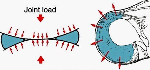
In effect, the meniscus creates stability and protects the underlying cartilage. Even partial meniscectomy (removing part of the meniscus) increases the risk of knee osteoarthritis by about 5 fold.
The blood supply of the meniscus is very important to understand, because it comes from the outside in. Meniscal tears in the outer third have the best chance of healing, while tears along the inner rim have a poor chance. This is critical to whether a tear is appropriate for suture repair (good blood supply) or partial meniscectomy and debridement (poor blood supply).
Symptoms & mechanisms
How meniscal tears happen, and how they feel
Acute meniscus tears can occur in younger patients with compression and rotation, but they are unusual in isolation: many occur together with other ligament injuries. Much more commonly we see chronic meniscal tears, which are more appropriately termed wear than tear, and occur with ageing. They are usually part of the spectrum of osteoarthritis, and surgery for these has been shown to be unhelpful in most cases.
Patients will generally have pain that localises to the side of their meniscal tear, swelling of the joint, and pain with loading or twisting movements.
When to seek a surgical opinion: whether acute or chronic, if you are having mechanical symptoms of locking, painful clicking, or giving way of the knee, a surgical opinion should be obtained.
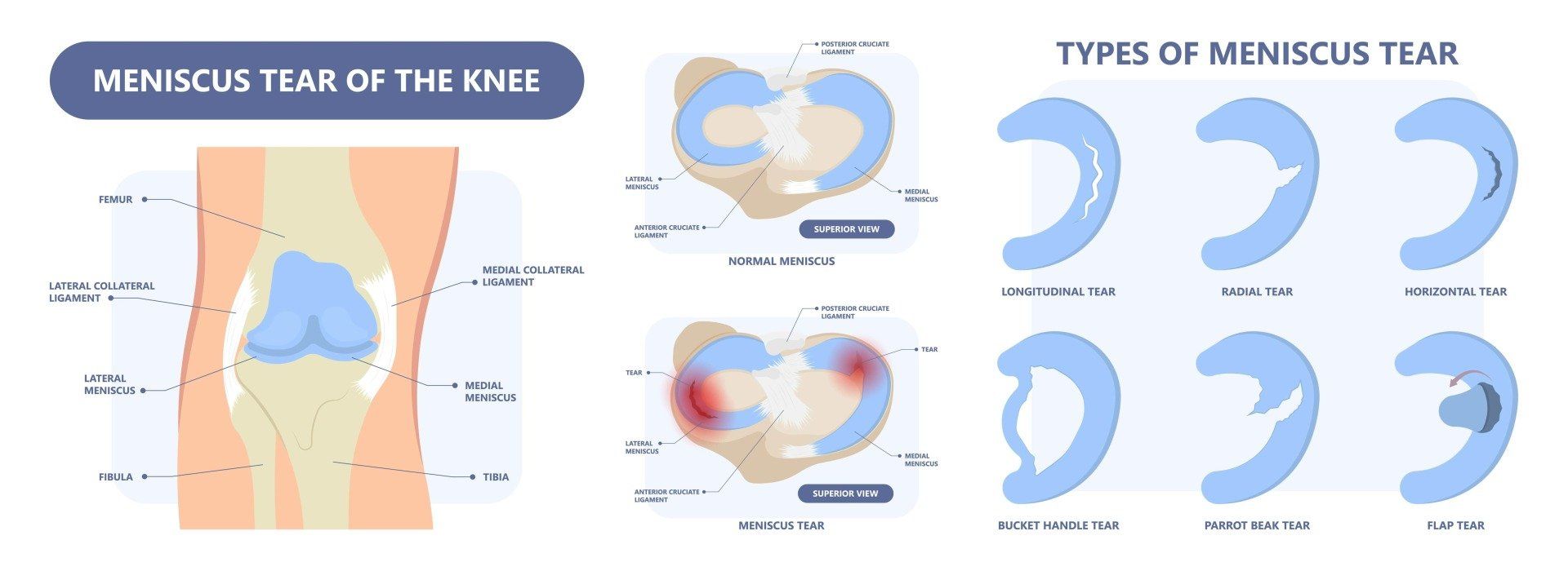
Peripheral tears occur toward the areas with more blood supply; when painful or unstable, a surgical opinion should be obtained. Tears can progress to unstable bucket handle tears, where a section flips in toward the middle of the joint. Radial tears involve the inner margin, which has a poor blood supply (a parrot beak tear is an extension of this). Complex tears combine orientations, usually affecting the inner avascular margin. Horizontal, degenerative tears occur as part of tissue wear; they can cause pain but are not repairable, and sometimes joint fluid leaks through the tear plane to gradually form a perimeniscal cyst (see the video animation below).
 Meniscal tears & perimeniscal cyst animation ▶ Watch on YouTube
Treatment
Treatment options for meniscal tears
The first line treatment for stable meniscal tears that aren’t displaced and causing locking of the knee is a non-surgical approach: relative rest, optimal loading, and sometimes anti-inflammatory treatments.
When pain and swelling impede even basic strength exercises, a short course of oral anti-inflammatories or the use of injection adjuncts can be very effective to settle pain and create an opportunity to redevelop strength. If cortisone is used, it should be used sparingly as a one-off injection: repeated injections in short succession have been shown to accelerate cartilage loss. Hyaluronic acid is an alternative adjunct for the osteoarthritic knee, and injections are delivered under ultrasound guidance for accuracy.
Biomechanical assessment and awareness of aggravating movements is very helpful to maximise recovery. After a minimum 8-week period of this approach, a surgical opinion is warranted if symptoms persist.
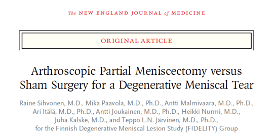
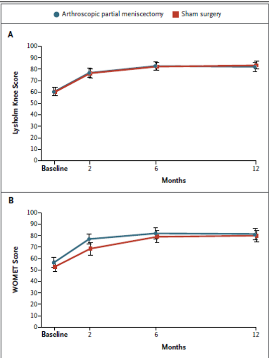
Multiple, consistent modern studies show no overall difference between arthroscopic surgery, sham surgery, or structured exercise for patients with stable degenerative meniscal tears. It is now standard of care to recommend a non-operative approach in the first instance.
Of course, trials tell us about the average and not the individual response, so this always needs to be an individual decision made with you.
The shoulder
Labral tears of the shoulder (glenoid labrum)
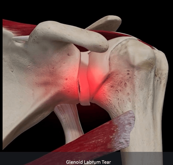
The glenoid labrum is a rubbery tissue that lines the shoulder socket like a white washer and deepens it, improving stability. The shoulder is a unique joint: inherently mobile at the expense of stability.
The labrum can be torn acutely with an instability episode of the shoulder, as shown in the video below. But it can also develop chronic tears from gradual tissue overload, especially in people with hypermobile joints that overstretch tissues easily.
 How a shoulder labral tear happens ▶ Watch on YouTube
The biceps tendon attaches to the top part of the labrum and acts as an important secondary stabiliser, strapping the ball down into the socket. During throwing or climbing, the tendon can pull forcefully at the labrum; if the labrum peels away from the socket, it can be painful every time the biceps is loaded.
 The biceps anchor & the labrum in detail ▶ Watch on YouTube
Chronic labral tears are a difficult and controversial area of sports medicine, because many patients have these tears but no pain. A tear may only hurt when it is repetitively loaded, or when it is very unstable and moving around.
Common signs include persistent deep-seated shoulder pain, clicking and clunking, and a sense of instability and distrust of the shoulder. Clinical tests are not very reliable, but an MRI with contrast (an arthrogram), often combined with a steroid injection for pain relief, picks up about 90% of labral tears while providing a platform of relief for rehabilitation exercise.
Once torn, the shoulder loses its snug fit and seal, allowing micro-movement. This can lead to pain from secondary sources such as bursitis or rotator cuff irritation as the ball wobbles in its socket. In chronic tears, especially with hypermobile shoulders, a period of rehabilitation of 4–6 months is warranted before considering surgical options, with activity demands strongly shaping the decision.
The hip
Labral tears of the hip (acetabular labrum)
The labrum of the hip, known as the acetabular labrum, is a biological washer that improves the congruence and stability of the joint. One of the most common reasons it tears is the shape of the hip joint itself lending toward abrasion of the labrum.
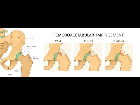
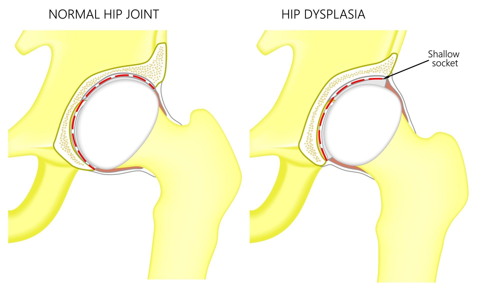
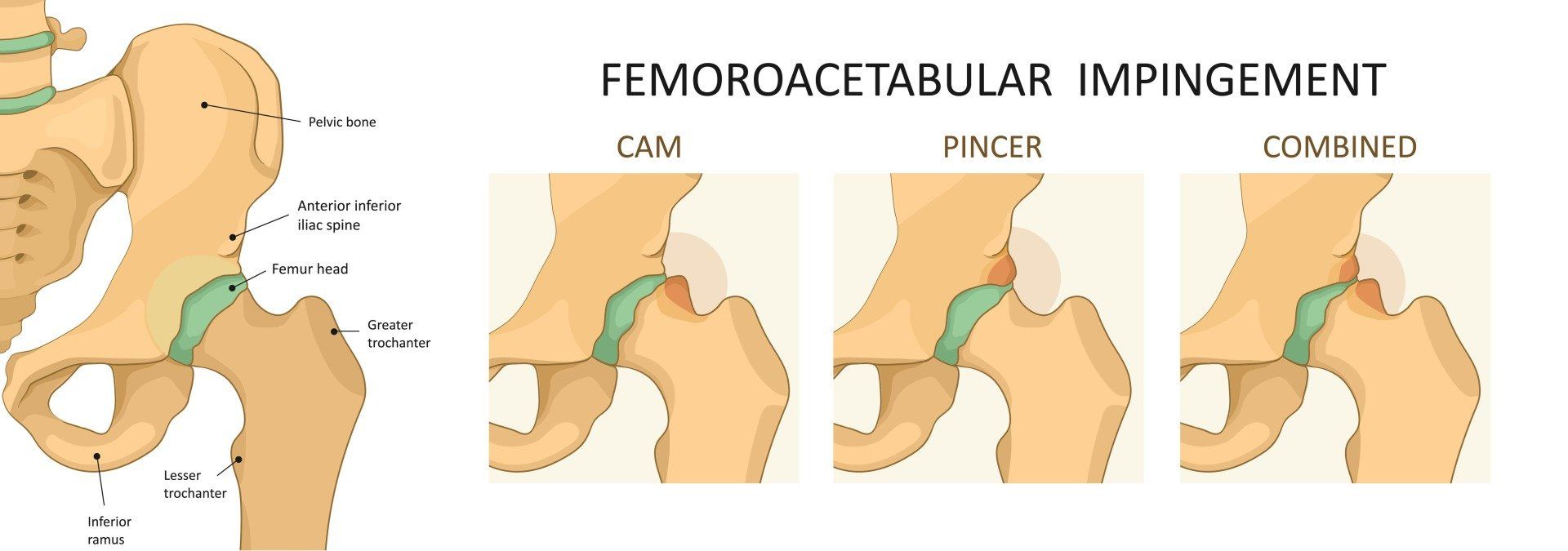
Some patients are born with a shallower hip socket (developmental dysplasia of the hip), which increases load on the edge of the socket. Others develop bony bumps around their growth plates during adolescence, leading to pinching (impingement) of the soft tissues at the front of the socket, including the labrum, as the knee moves toward the chest. This is called FAI or hip impingement syndrome; you can read more on our hip & groin pain page.
A challenge for clinicians is that labral tears are not always painful. One prospective study showed 40% of patients with a symptomatic hip labral tear had a completely asymptomatic tear on the other side, and as many as 55% of athletes meet imaging criteria for FAI, yet only a small proportion have FAI syndrome.
The evidence
Key references
The practice-changing studies behind the recommendations on this page:
- Sihvonen R, et al. Arthroscopic partial meniscectomy versus sham surgery for a degenerative meniscal tear. N Engl J Med 2013;369:2515–2524. doi:10.1056/NEJMoa1305189
- Katz JN, et al. Surgery versus physical therapy for a meniscal tear and osteoarthritis (METEOR). N Engl J Med 2013;368:1675–1684. doi:10.1056/NEJMoa1301408
- Papalia R, et al. Meniscectomy as a risk factor for knee osteoarthritis: a systematic review. Br Med Bull 2011;99:89–106. doi:10.1093/bmb/ldq043
- Zeng C, et al. Intra-articular corticosteroids and the risk of knee osteoarthritis progression: results from the Osteoarthritis Initiative. Osteoarthritis Cartilage 2019;27:855–862. doi:10.1016/j.joca.2019.01.007
- Frank JM, et al. Prevalence of femoroacetabular impingement imaging findings in asymptomatic volunteers: a systematic review. Arthroscopy 2015;31:1199–1204. doi:10.1016/j.arthro.2014.11.042
Common questions
Meniscus & labral tear FAQs
Usually not. For stable tears that are not causing locking of the knee, multiple modern trials, including a landmark study comparing real surgery with sham surgery, show no overall benefit from an operation. Non-surgical care is now the standard first-line approach, with a surgical opinion warranted if a proper trial of at least 8 weeks does not settle symptoms.
Mechanical symptoms: locking of the knee, painful clicking, or giving way. These suggest an unstable tear, such as a displaced bucket handle tear, where surgery has a clearer role. Painful or unstable peripheral tears, which sit in the zone with better blood supply, may also be suitable for repair.
Yes, when pain and swelling are blocking even basic strength work, an injection adjunct can settle the joint and open a window for rehabilitation. If cortisone is used it should be a sparing, one-off injection, because repeated injections in short succession have been shown to accelerate cartilage loss. Hyaluronic acid is an alternative for the osteoarthritic knee.
No, and this is one of the most important ideas on this page. Many people have labral tears with no pain at all: in one study, 40% of patients with a painful hip labral tear had a completely silent tear on the other side, and as many as 55% of athletes meet imaging criteria for hip impingement while only a small proportion have symptoms. We treat the person and their examination findings, not the scan alone.
For stable knee meniscal tears, a minimum of 8 weeks of structured non-surgical care before seeking a surgical opinion. For chronic labral tears of the shoulder, especially in hypermobile patients, a period of 4 to 6 months of rehabilitation is warranted before considering surgical options, with your activity demands strongly shaping the decision.
A tear on your scan, or a knee, shoulder or hip you don’t trust?
Our Sport and Exercise Physicians in Leichhardt can work out whether the tear is actually the cause of your pain, exclude the problems that genuinely need a surgeon, and build an evidence-based rehabilitation plan first.
This page provides general information only and is not a substitute for personalised medical advice. Please consult a qualified health practitioner for assessment of your individual circumstances.