


Knee Conditions • Progressive Sports Medicine
Runner’s Knee (Patellofemoral Pain Syndrome)
A dull ache under and around the kneecap that flares with stairs, squatting, lunging and sitting too long. It is common, it is treatable, and it very rarely needs surgery.
Introduction
What is Runner’s Knee?
Runner’s Knee is similar but different to Jumper’s Knee. They can co-exist.
The medical term for Runner’s Knee is Patellofemoral Pain Syndrome. This refers to pain under and around your kneecap.
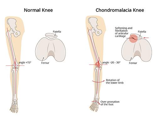
▶ Watch: Patellofemoral Pain Syndrome explainedRunner’s Knee has a number of synonyms, including anterior knee pain syndrome, patellofemoral maltracking, movie-goer’s knee and chondromalacia patella. All refer to pain generated by anatomical structures around the joint between your kneecap (patella) and the groove formed by the lower part of the thigh bone (trochlea).
As the name suggests, Runner’s Knee is a common complaint among runners, jumpers, and other athletes such as skiers, cyclists and soccer players.
Anatomy
The patellofemoral joint
The patellofemoral joint is the joint between the kneecap and the groove that is formed at the end of the thigh bone, called the trochlea.
This joint enjoys the thickest articular cartilage in the body, and it needs it: the cartilage must absorb both compressive and shear forces. Every time you bend your knee, the cartilage surfaces engage at about 30 degrees of knee bend.
10–12× body weight
When you lunge or go up or down stairs, the patellofemoral joint must absorb ten to twelve times your body weight in force.
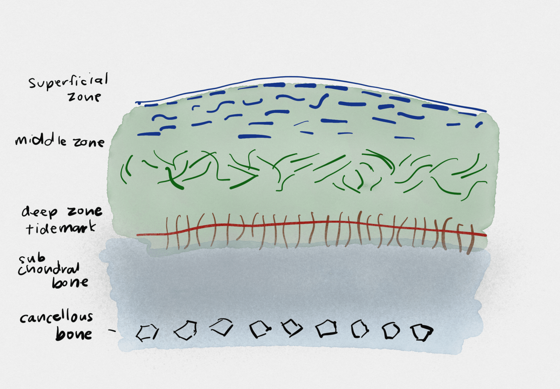
Hyaline cartilage has a very unique architecture. The top layers have collagen fibres oriented horizontally to resist shear forces, while the deeper layers have the collagen pointing vertically to absorb the compressive and impact forces.
When the kneecap does not track smoothly in its groove, it can lead to increased shear forces that abrade and irritate the cartilage. Even though there is no deep or significant damage, the excessive friction on the superficial layers (see the cartilage image below) can lead to pain, swelling and stiffness that often limits exercise capacity and persists for months.
Causes
Why Runner’s Knee develops
Runner’s Knee can result from poor alignment of the kneecap, complete or partial dislocation, overuse, tight or weak thigh muscles, flat feet, or even direct trauma to the knee.
It is important to realise that this is more of an inflammatory response, where the knee joint lining detects small, microscopic cartilage fragments, rather than any significant visible cartilage damage. Patellofemoral pain often comes from inflamed soft tissues (fat, ligaments and capsular tissue) and irritation or softening of the cartilage that lines the underside of the kneecap.
Cartilage itself has no nerve endings, so we know it does not generate the pain. The underlying bone, however, has many nerve endings, and becomes exposed when the cartilage thins or is injured.
Pain in the knee may also be referred from, or contributed to by, other parts of the body, such as the feet, back or hip.
Symptoms
What Runner’s Knee feels like
The classic symptom is a dull aching pain underneath the kneecap while doing anything that loads the patellofemoral joint, including walking up or down stairs, squatting, lunging or kneeling.
One of the old names for the condition is “movie-goer’s knee”, because a common complaint is that pain is worse after standing up from prolonged sitting.
Diagnosis
How we diagnose Runner’s Knee
Most of the features of patellofemoral pain syndrome are clinical and functional, because it is a dynamic process. A classical history of pain, the presence of known risk factors, pain during a squat movement, tenderness of the edges of the patella and a grinding sensation during movement all support the diagnosis.
Diagnoses to exclude
The major diagnoses to exclude are instability of the kneecap (that is, dislocations) and a condition called excessive lateral pressure syndrome, a much more severe form of mechanical overload of the outer part of the patella due to malalignment. That condition often requires surgery, while Runner’s Knee does not.
Imaging
Imaging findings such as a high-seated kneecap or thinning of the cartilage on MRI also support the diagnosis of patellofemoral maltracking, but they are not essential for diagnosis.
Dr Samra and our Sport and Exercise Physicians will assess your specific risk factors and contributors, including your running and walking biomechanics and any “weak links” in the chain from top to bottom.
Risk factors
Who is at risk?
There are many biomechanical, anatomical and load-related factors that predispose a person to patellofemoral pain syndrome:
- A kneecap that sits higher, or a smaller kneecap
- A flatter groove for the kneecap, called trochlea dysplasia
- Alignment that tends to make the patella shift or tilt to the outer side of the knee, increasing the contact forces on this area
- Females are at higher risk
- Poor stabiliser muscle activity around the knee and hip
- Short hamstrings and weak quadriceps muscles
Treatment
First response: settle the pain, keep moving
Treatment of Runner’s Knee can begin immediately after the injury is sustained. Nonsteroidal anti-inflammatory medications (NSAIDs, such as ibuprofen or naproxen) can relieve some of the pain or discomfort. Alongside this, we follow the POLICE principles to reduce acute pain and swelling:
- P
Protect: support and position the injury in a way that doesn’t worsen it.
- OL
Optimal Loading: rehabilitation begins as soon as the injury occurs. It is a matter of finding non-exacerbating load to stimulate and enhance tissue recovery.
- I
Ice: for comfort and pain relief. 10 minutes, as often as every 4 to 6 hours, for the first 2 days.
- C
Compression: to control swelling and inflammation.
- E
Elevation: the most potent means of reducing swelling is lifting the limb above the level of the heart.
Activity modification: pause aggravating athletic activities until symptoms have reduced or resolved, then reintroduce load gradually with pacing to find the right dose for the joint during recovery.
Treatment
Recommended treatments for Runner’s Knee
The best consensus guidelines that summarise the evidence tell us that treatment for patellofemoral pain must be individualised, based on a full assessment of your specific contributors to pain. Even when there is marked wear of the cartilage and osteoarthritis of the patellofemoral joint, non-surgical treatments should be targeted first line, because they can make a big difference to pain.
Evidence shows that multimodal treatment programs lasting at least six weeks have a very significant benefit for pain and function.
These programs incorporate education about the pain and the most suitable activities, customised patella taping (which usually makes a significant difference), sometimes orthotics, and short courses of oral anti-inflammatories. All of this is designed to promote the most important aspects of long-term recovery: specific progressive resistance training of the quadriceps, stability exercises, and hamstring and quadriceps lengthening exercises.
Exercise training
Once pain is controlled, exercise to strengthen the stabilising muscles of the hip and knee can improve your exercise tolerance and reduce the symptoms of Runner’s Knee. Core stability (Pilates-style) exercises are often a missing link that helps people prevent pain coming back, by improving movement control and quality.
Taping
An adhesive tape is applied over the patella to alter the kneecap alignment and movement. Taping of the patella may reduce pain by more than 50% immediately.
Bracing
Your physician may recommend stabilising the patella with either a brace or athletic tape to keep it in place during exercise training.
Orthotics
Shoe inserts (over the counter or customised) may be prescribed for those with arches that fall in (pronate), and this can help relieve the pain.
Anti-inflammatory medications
NSAIDs used for a short 1 to 2 week period can reduce pain and swelling to promote rehabilitation.
Injection therapies
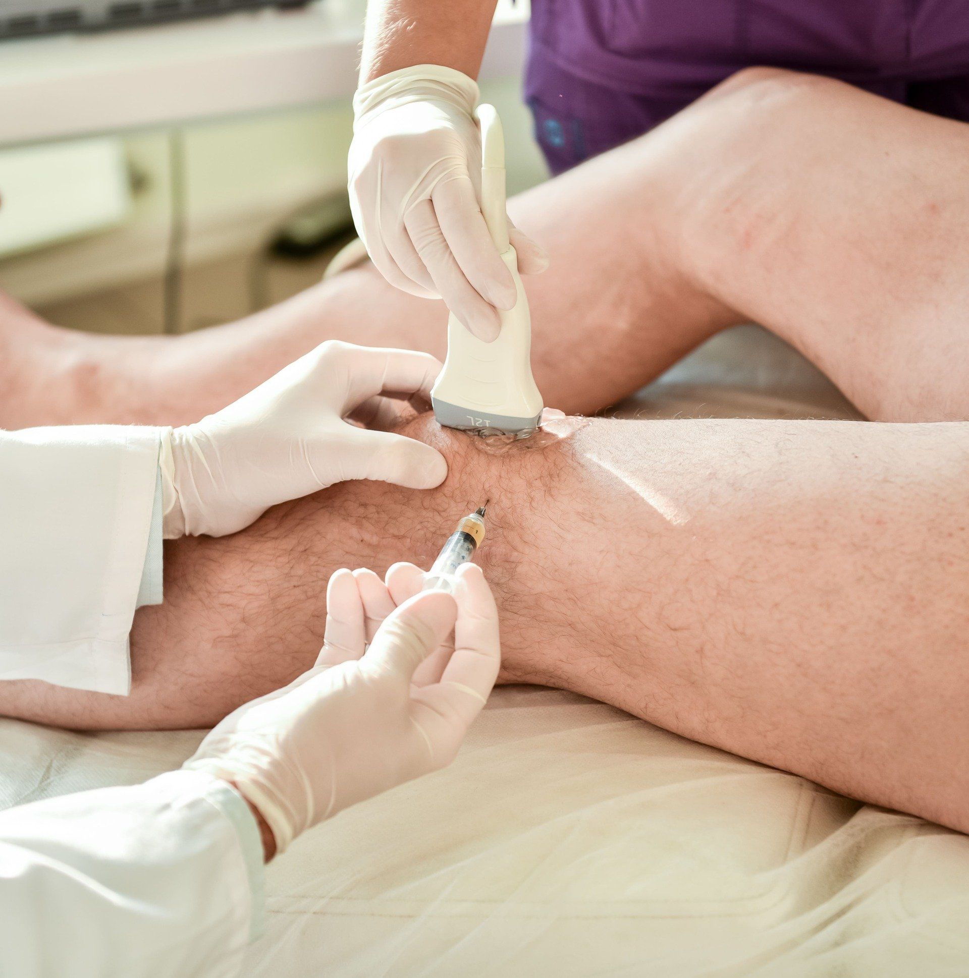
A single cortisone injection may be used to reduce pain and inflammation, particularly in recalcitrant cases where the fat pads around the knee are very sensitive or inflamed. It is only used to provide a window of opportunity, so that your own stabilising muscles can be retrained; pain inhibits these muscles and perpetuates a maltracking patella. Injections are delivered as ultrasound-guided injections for accuracy.
Platelet-rich plasma (PRP) therapy
This practice, also known as PRP, involves injecting the site of the injury with the patient’s own platelet-rich plasma, in an effort to accelerate healing.
Pain education matters. Approaching pain from a biopsychosocial model is important in addressing fear of movement that comes from unhelpful thinking, such as the belief that pain means tissue damage. The body is very resilient and dynamic. It is constantly healing and adapting to microscopic damage, and exercise helps promote this adaptation, so long as it does not exacerbate pain.
Additional treatments
Surgery: rarely needed
In some rare cases, surgery may be needed. Options include:
Arthroscopy
A small camera and several surgical tools are inserted into the knee joint and used to remove small fragments or damaged tissue. Some patients have an extra fold of joint tissue called a plica that rubs on the inside of the kneecap joint, and this can be shaved.
Realignment surgery
Used to realign the patella and reduce the abnormal pressure on cartilage and the supporting structures around the front of the knee. There are a number of options, depending on the specific cause of maltracking.
For Runner’s Knee, surgery is the exception, not the rule. Non-surgical treatment is first line, and most people never need an operation.
Prevention
Preventing Runner’s Knee
- If you are overweight, you may need to control your weight to avoid overstressing your knees.
- Gradually increase the intensity of your workouts, and avoid running on hard surfaces without adequate conditioning.
- Wear properly fitting, good quality running shoes with good shock absorption.
- Avoid running straight down hills. Instead, walk down or run in a zigzag pattern.
- If in doubt, speak to your physiotherapist and sit down to plan a regular program that promotes strength and control exercises. If you have not tried customised taping as a starting point, this should be explored.
- Core stability exercise is often used to treat back pain. It is under-rated in preventing upper and lower body injuries.
- Programs such as the FIFA 11+ provide a good starting point, as evidence-based warm-ups that have been shown to lower injury risk.
Common questions
Runner’s Knee FAQs
No, though they can co-exist. Runner's Knee (patellofemoral pain syndrome) is a diffuse ache under and around the kneecap from the joint between the patella and the thigh bone. Jumper's Knee is a patellar tendon problem, with focal tenderness just below the kneecap that you can point to with one finger.
Usually not. Cartilage has no nerve endings, so it does not generate the pain. The pain typically comes from inflamed soft tissues and irritation of the joint lining, often with only superficial cartilage changes. Pain does not equal tissue damage, and understanding this is itself part of effective treatment.
Very rarely. The evidence strongly supports non-surgical treatment first line, even when there is cartilage wear or patellofemoral osteoarthritis. Surgery is reserved for specific problems such as recurrent dislocation or severe malalignment, which are excluded during your assessment.
Multimodal programs lasting at least six weeks show a very significant benefit for pain and function. Many people improve steadily over two to three months as strength and movement control build, and continuing the exercises protects against recurrence.
Sometimes, in recalcitrant cases, particularly where the fat pads around the knee are very sensitive or inflamed. A single injection is used only to open a window of opportunity for retraining your stabilising muscles, and it is delivered under ultrasound guidance for accuracy.
Kneecap pain slowing you down?
Our Sport and Exercise Physicians in Leichhardt can assess your knee and biomechanics, exclude the conditions that behave differently, and build an individualised program to get you moving comfortably again.
This page provides general information only and is not a substitute for personalised medical advice. Please consult a qualified health practitioner for assessment of your individual circumstances.